Sutthiwan Thammawat1 ![]() ,
Kusavadee Sangdee1,
Aphidech Sangdee2,3
,
Kusavadee Sangdee1,
Aphidech Sangdee2,3
For correspondence:- Sutthiwan Thammawat Email: tham.sutthi76@gmail.com Tel:+6686-9890776
Received: 22 October 2016 Accepted: 24 January 2017 Published: 25 February 2017
Citation: Thammawat S, Sangdee K, Sangdee A. Time-kill profiles and cell-surface morphological effects of crude Polycephalomyces nipponicus Cod-MK1201 mycelial extract against antibiotic-sensitive and -resistant Staphylococcus aureus. Trop J Pharm Res 2017; 16(2):407-412 doi: 10.4314/tjpr.v16i2.20
© 2017 The authors.
This is an Open Access article that uses a funding model which does not charge readers or their institutions for access and distributed under the terms of the Creative Commons Attribution License (http://creativecommons.org/licenses/by/4.0) and the Budapest Open Access Initiative (http://www.budapestopenaccessinitiative.org/read), which permit unrestricted use, distribution, and reproduction in any medium, provided the original work is properly credited..
Purpose: To examine the effect of crude Polycephalomyces nipponicus Cod-MK1201 mycelial extract on the viability and cell surface morphology of methicillin-susceptible Staphylococcus aureus (MSSA) and methicillin-resistant Staphylococcus aureus (MRSA).
Methods: Time-kill assays were conducted by incubating test bacteria with the extract and sampling at selected time points within a 24 h period. The effects of the extract on MSSA and MRSA ultrastructure were determined using a scanning electron microscope (SEM).
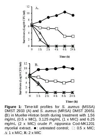
Results: Time-kill assay data indicate a bactericidal effect against both strains of staphylococci. The extracts were rapidly bactericidal at concentrations of 1 x MIC and 2 x MIC, achieving complete elimination of the test bacterial strains within 2 h. SEM micrographs of S. aureus taken after treatment with various concentrations of the extract revealed extensive morphological alterations to the cell surface of both MSSA and MRSA.
Conclusion: The results confirm the antibacterial activity of P. nipponicus Cod-MK1201 mycelial extract. Further research may allow this to be developed as an alternative therapy to alleviate S. aureus infection
Introduction
Staphylococcus aureus is a major human pathogen, causing pyogenic infections such as abscesses, and even fatal septicemia [1]. Methicillin-resistant S. aureus (MRSA) infections have become an important problem in both the hospital and community requiring vancomycin for effective clinical treatment [2,3]. Alarmingly, MRSA strains with reduced susceptibility to vancomycin are now also being reported [4,5]. Moreover, there have been reports of MRSA resistant to chlorhexidine and other disinfectants commonly used for infection control and reducing the spread of MRSA in hospitals [6-8]. To overcome the problem of antibiotic resistance, alternative sources of bioactive compounds are being screened for possible development as the next generation of antibacterial therapies.
Medicinal mushrooms have been used as traditional remedies for infectious diseases in many tropical countries, and represent a natural source of antimicrobial agents [9]. Cordyceps is a genus of entomopathogenic fungi that are used in traditional Chinese medicine for various health effects including immunomodulatory, anticancer, antioxidant, anti-inflammatory and antimicrobial activities [10-13]. Recently, it was demonstrated that Cordyceps taii extracts have broad-spectrum activity against both bacteria and fungi [14]. Also, a protein isolated from the cultured mycelia of Cordyceps sinensis has been reported to have activity against the bacteria Staphylococcus aureus, Escherichia coli, Proteus vulgaris, , and Salmonella enterica serovar Typhi [15]. The methanol extract from Cordyceps militaris has strong antibacterial and antifungal activity too [10]. These findings indicate that many entomopathogenic fungi produce bioactive compounds effective against microbial pathogens. However, time-kill data and information on the morphological effects of these extracts on bacterial cells is limited.
In a previous study, we isolated the entomopathogenic fungus Cod-MK1201 from a dead cicada nymph and found it to have potent antibacterial activity against both Gram-positive and Gram-negative bacteria [16]. The isolate was subsequently identified as Polycephalomyces nipponicus based on fungal morphology, three regions of ribosomal nuclear DNA (ITS, LSU, SSU), and four protein-coding regions (rpb1, ef1α, β-tubulin, ATP6) [17]. However, the bactericidal activity of the crude extract of this isolate has not yet been characterized in terms of the kinetics of bacterial death. Therefore, the aim of the present study was to evaluate the activity of Cod-MK1201 ethanol extract against Gram-positive Staphylococcus aureus by the time-kill method. The effect of the ethanol extract on bacterial morphology was also examined.
Methods
Microorganisms and antibacterial agent
Two reference strains of Staphylococcus aureus, methicillin-sensitive Staphylococcus aureus (MSSA) strain DMST 2933 and methicillin- resistant Staphylococcus aureus (MRSA) strain DMST 20651 were obtained from the medical microorganism collection at the Department of Medical Sciences Thailand (DMST), Ministry of Public Health, Thailand. These were used in assays evaluating the antibacterial activity of the extract.
Preparation of mycelial extract of Polycephalomyces nipponicus Cod-MK1201
Mycelial extracts were prepared as described previously by Sandgee et al [16]. Briefly, mycelial discs were cut and inoculated into 25 ml of induced medium, then incubated at 28 °C with shaking. After 20 days, the mycelium on the surface of the culture medium was collected, dried (50 °C overnight), powdered, and stored in a screw cap tube. Ethanol 50 % (v/v) was then added to the dried mycelium, and the mycelial suspension (100 mg/ml) was sonicated with a high Intensity Ultrasonic Processor (Model VCX750, Newtown, CT, USA). This step was performed on ice as described by Sangdee et al [18].
Time-kill assay
Time-kill assays were performed as described by White et al [19] and Aiyegoro et al [20], with some modifications regarding the counting of viable cells. MICs were determined by broth microdilution assay as described by Sangdee et al [16]. The extract was incorporated into 0.9 mL Mueller Hinton broth (MHB) at concentrations of 1.56 mg/mL (0.5 x MIC), 3.125 mg/mL (1 x MIC) and 6.25 mg/mL (2 x MIC). Test tubes of MHB without extract were used as growth controls. Overnight cultures of the bacterial strains at cell densities of approximately 1 x 10⁸ CFU/mL were used to inoculate 0.1 mL volumes of both test and control tubes. The cultures were then incubated in a shaker at 37 °C for 1, 2, 4, 6 and 24 h. After each interval, ten-fold serial dilutions were prepared with phosphate buffered saline (PBS), and 0.1 mL samples were pipetted onto Mueller Hinton agar (MHA) plates in duplicate. Colony counts were performed after 18 h incubation at 37 °C. Plates with 30–300 colonies were used for these counts, and the kill rate was determined by plotting log10 viable counts (CFU/mL) against time. Bactericidal activity was defined as a ≥ 3 log10 decrease in CFU/mL of the initial microbial population, while bacteriostatic activity was defined as a <3 log10 decrease in CFU/mL.
Scanning electron microscopy (SEM)
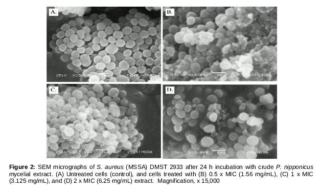
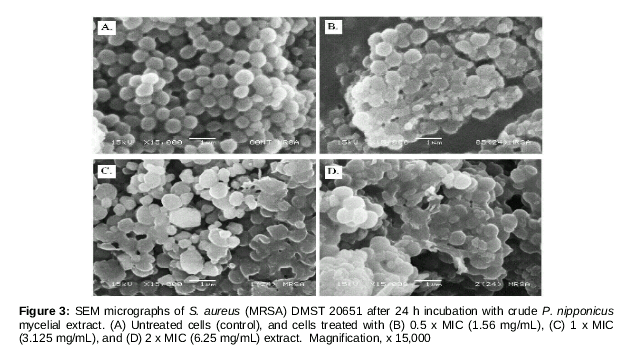
Scanning electron microscopy was used to determine the antibacterial effect of the mycelial extract on S. aureus cells. Briefly, the test bacteria were cultured in MHB and incubated at 37°C with shaking for 3 h. The turbidities of the final inocula were adjusted to the 0.5 McFarland standards. One hundred microliters of each bacterial cell suspension was then added to 900 µL of the mycelial extract (at concentrations of 0.5 x MIC, 1 x MIC or 2 x MIC) in MHB. Untreated cells were used as growth controls. The cultures were then incubated in a shaker at 37°C for 24 h. Each sample was placed in a separate vial, and cells from each tube were harvested by centrifugation. A 2.5 % glutaraldehyde solution was used to prefix all samples overnight at 4 °C. Thereafter, the samples were rinsed 3 times with PBS, and post-fixed in 1 % osmium tetroxide in the same buffer for an hour. This was followed by 4 further rinses with PBS. After fixation, a graded series of acetone was used to dehydrate the specimens with 100 % acetone being used for the final step. The specimens were then mounted and coated with gold before examination by scanning electronic microscopy (JOEL Tsm-700 FSHL, Japan).
Results
Time-kill profiles
Time-kill assays allow antibacterial agents to be classified as bacteriostatic or bactericidal, and characterization of the relationship between agent concentration and activity over time. The results obtained with the mycelial extract and two S. aureus strains (MSSA strain DMST 2933 and MRSA strain DMST 20651) are shown in . The increase in viable count of bacteria in the control group shows these bacteria were actively growing from 1 to 24 h. After 24 h incubation with 0.5 x MIC (1.56 mg/mL) of the extract, a 3 log10 CFU/mL reduction in viability of S. aureus DMST 2933 occurred (A), indicating the extract is bactericidal against this strain of MSSA. Comparable results were not obtained with 0.5 x MIC extract and S. aureus DMST 20651 (B), indicating this concentration of extract is bacteriostatic against MRSA.
At a concentration of 1 x MIC (3.125 mg/mL), however, the extract was bactericidal against both tested organisms by 24 h. Also, increasing the concentration to 2 x MIC (6.25 mg/mL) resulted in bactericidal activity against MSSA by 1 h (A). Complete elimination of both test strains was achieved by 2 h treatments with 1 x MIC and 2 x MIC extract. These results indicate that bactericidal activity of the extract is both time- and concentration-dependent.
Morphological characteristics
Samples of P. nipponicus mycelial extract-treated S. aureus were examined by SEM to detect any physical changes in appearance of the cells. SEM micrographs of S. aureus after 24 h treatment with various concentrations (0.5 x MIC, 1 x MIC and 2 x MIC) of the extract revealed considerable morphological alterations to both the MSSA and MRSA strains ( and ). shows the SEM images of treated and untreated control samples of S. aureus (MSSA) DMST 2933. Untreated cells were intact, smooth, and of normal coccal morphology (A). The destructive effects of the extract on test bacteria can be seen in B-D. Bulges are present at the cell surface and debris is observed, much more so than in the untreated control cells.
Characteristic morphological alterations observed in S. aureus (MRSA) DMST 20651 treated with mycelial extract are shown in . Exposure to 0.5 x MIC, 1 x MIC and 2 x MIC of the mycelial extract for 24 h led to damage of the cell wall (B-3D), as indicated by bulging at the cell surface. Untreated control cells, by comparison, had a smooth surface (A). When MRSA was treated with 6.25 mg/mL (2 x MIC) of the extract, cell morphology and surface appearance were dramatically altered (D). Whilst the samples were not prepared in a quantitative manner, it is clear from the micrographs that the number of damaged cells in the treated vials greatly outnumbers those in the control vials. These results indicate that the active compound had some effect on the cytoplasmic membrane and cell wall of bacteria. This could have been caused by an alteration in cytoplasmic membrane permeability, leading to cell wall leakage.
Discussion
Increases in the incidence of multidrug-resistant S. aureus infections have prompted renewed efforts to identify biologically active molecules from natural sources that might be useful antibacterial therapies. Several Cordyceps species extracts, including those from C. sinensis [15], C. militaris [10], C. sobilifera [9], and C. taii [14], have been reported to have potent antibacterial activity against human pathogenic bacteria. In a previous study, we demonstrated the antibacterial effects of an ethanolic mycelial extract of P. nipponicus Cod-MK1201 against MSSA and MRSA strains. Because the MIC and MBC values of the extract were quite low (3.125 mg/mL) [16], it was decided that further characterization would be useful. Therefore, the present study determined the time-kill profiles and morphological effects of the extract against MSSA and MRSA. The findings obtained represent the first published information on the microbial death kinetics and possible mechanism of action of P. nipponicus mycelial extract against antibiotic -susceptible and -resistant strains of S. aureus.
The time-kill assay is an important method for determining the potency of bioactive extracts, and determining whether these are bactericidal or bacteriostatic. The present study analyzed bacterial viability over time during treatment with various concentrations of P. nipponicus mycelial extract. At sub-inhibitory levels (0.5 x MIC), the extract had bactericidal activity against MSSA, and bacteriostatic activity against MRSA. The mycelial extract at a concentration equal to 2 x MIC was rapidly bactericidal, achieving complete elimination of both test bacterial strains within 2 h. All of the time-kill data obtained with the P. nipponicus mycelial extract showed its antibacterial activity to be time - and concentration-dependent. This finding correlates with that of Okemo et al [21], who found that Ximenia caffra extracts kill S. aureus in both a time- and concentration-dependent manner. Also, extracts of the endophytic fungus Nigrospora sphaerica CL-OP30 are active against S. aureus in a time- and concentration-dependent manner during the first 12 h of treatment [22].
In addition to the time-kill data obtained, the effect of the P. nipponicus mycelial extract (0.5 x MIC, 1 x MIC and 2 x MIC) on S. aureus morphology was determined by SEM. Perturbations to cell surface morphology could be detected when MSSA and MRSA were treated with various concentration of the mycelial extract. Scanning electron microscopy images captured at 15,000x magnification clearly show cell surface and morphological changes such as bulges, surface disruption, and broken cells. These alterations may be due to phenolic or other bioactive compounds present in the mycelial extract affecting the cell wall and cytoplasmic membrane of S. aureus, thereby causing leakage of bacterial cell contents and eventual cell lysis [16,23]. Similar results have been described in a study where S. aureus cells were treated with Nigrospora sphaerica CL-OP30 extract. The authors attributed the formation of cavities, surface alterations, and appearance of collapsed and shrunken cells to leakage of cytoplasmic contents [22]. Similar cell wall effects have been reported in S. aureus treated with bacteria-derived antibiotics [24,25].
Conclusion
The results of this study reveal that P. nipponicus Cod-MK1201 mycelial extract exhibits significant in vitro antibacterial activity against staphylococci. These entomopathogenic fungi may therefore be suitable candidates for bioprospecting for antimicrobial drugs. Further studies will be required to isolate and identify the bioactive compounds present in P. nipponicus Cod-MK1201, and determine whether they could be developed as novel drugs for the treatment of antibiotic-resistant bacterial infections.
Declarations
Acknowledgement
References
Archives


News Updates